Skip to content
登入/註冊
食品所
舊網站
Search
Youtube
Map-marked-alt
Choose a language
中文 (台灣)
English
首頁
關於生資中心
中心簡介
中心組織
中心沿革
創新研發與應用
生物材料寄存及分讓辦法
公正性聲明
關鍵技術
生物資源之培養與保存技術
生物資源之開發與應用技術
對外服務
生物資源提供
生物資源引進
委託試驗
菌種鑑定與人類細胞複核
寄存服務
秘密寄存
公開寄存
專利寄存
農藥微生物種原寄存
委託代訓
最新消息 & 網頁新知
出版品
生資中心簡訊
其他出版品
相關聯結
生物資源線上目錄
BCRC客戶服務系統
微生物條碼資料庫系統
BCRC小教室
專利生物材料服務e平台
台灣微生物知識網
國家衛生研究院細胞庫
產業服務平台
TSCB台灣幹細胞庫
新資源
NBRC-BCRC 國際合作專區
聯絡我們
FAQs
同仁專區
BCRC工作流程管理系統
生物資源管理系統
Menu
首頁
關於生資中心
中心簡介
中心組織
中心沿革
創新研發與應用
生物材料寄存及分讓辦法
公正性聲明
關鍵技術
生物資源之培養與保存技術
生物資源之開發與應用技術
對外服務
生物資源提供
生物資源引進
委託試驗
菌種鑑定與人類細胞複核
寄存服務
秘密寄存
公開寄存
專利寄存
農藥微生物種原寄存
委託代訓
最新消息 & 網頁新知
出版品
生資中心簡訊
其他出版品
相關聯結
生物資源線上目錄
BCRC客戶服務系統
微生物條碼資料庫系統
BCRC小教室
專利生物材料服務e平台
台灣微生物知識網
國家衛生研究院細胞庫
產業服務平台
TSCB台灣幹細胞庫
新資源
NBRC-BCRC 國際合作專區
聯絡我們
FAQs
同仁專區
BCRC工作流程管理系統
生物資源管理系統
1
2
3
4
5
6
7
8
9
10
11
12
13
對外服務
菌株購買
多樣、本土、新穎性及應用性生物資源
More
委託試驗
符合國際標準及品質認證之測試試驗項目
More
技術發展
歷年投入之研究產出及可移轉技術及專利
More
菌種鑑定
微生物及細胞鑑定、複核及客製化服務
More
菌株寄存
公開、秘密、專利及農藥微生物等之寄存
More
即時訊息
最新消息
網頁新知
生資中心簡訊
最新消息
2026-03-12
115年清明節連假細胞株訂單收件截止日期更動通知
2026-03-12
2026 細胞培養與品管研習會 Cell Culture Workshop
2026-02-08
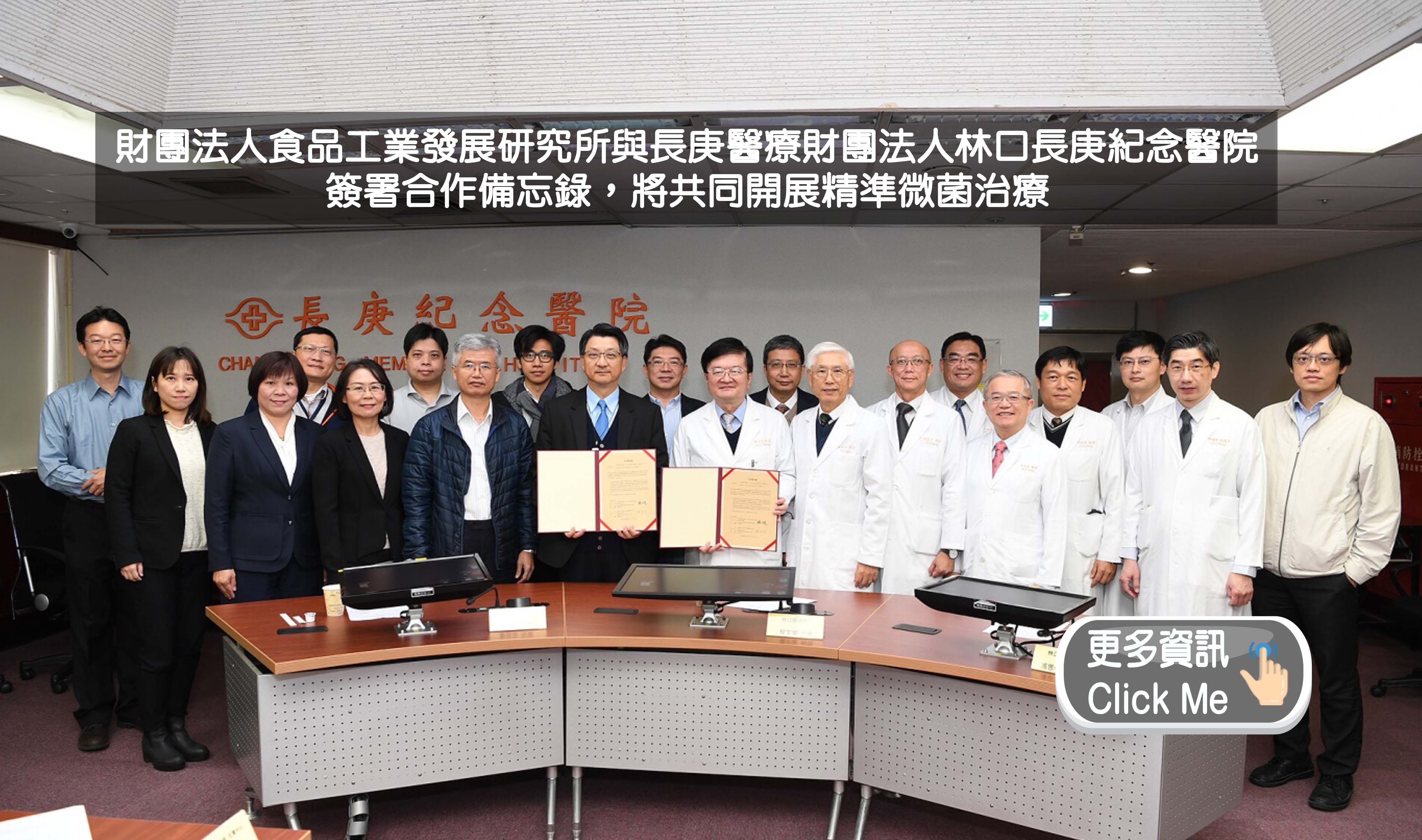
115年度經濟部智慧財產局指定專利生物材料之國內寄存機構為財團法人食品工業發展研究所
2026-01-13
2026年農曆春節期間生資中心暫停服務公告
2025-12-18
食品所生資中心獲 TAF 認證為【全國首家取得 ISO 20387 認證之生物資料庫】
+更多資訊
網頁新知
2026-03-19
微生物命名新篇章:從純培養到 SeqCode 的雙軌制與衝擊
2026-02-09
重塑永續未來的「奇蹟分子」-微生物界面活性劑之潛力與應用
2026-01-26
非侵入性大腸癌篩檢之新途徑
2026-01-12
大腦鋰缺乏可能導致阿茲海默症?
2025-12-28
以球形紅桿菌萃取物作為細胞培養系統替代補充劑的潛力
+更多資訊
生資中心簡訊
第136期簡訊
第135期簡訊
第134期簡訊
第133期簡訊
第132期簡訊
+更多資訊
推薦影音
Menu
首頁
關於生資中心
中心簡介
中心組織
中心沿革
創新研發與應用
生物材料寄存及分讓辦法
公正性聲明
關鍵技術
生物資源之培養與保存技術
生物資源之開發與應用技術
對外服務
生物資源提供
生物資源引進
委託試驗
菌種鑑定與人類細胞複核
寄存服務
秘密寄存
公開寄存
專利寄存
農藥微生物種源寄存
委託代訓
最新消息 & 網頁新知
出版品
生資中心簡訊
其他出版品
聯絡我們
FAQs
相關聯結
生物資源線上目錄
BCRC客戶服務系統
微生物條碼資料庫系統
BCRC小教室
專利生物材料服務e平台
台灣微生物知識網
國家衛生研究院細胞庫
產業服務平台
TSCB台灣幹細胞庫
新資源
NBRC-BCRC 國際合作專區
同仁專區
BCRC工作流程管理系統
生物資源管理系統
最新消息
2026-03-12
115年清明節連假細胞株訂單收件截止日期更動通知
2026-03-12
2026 細胞培養與品管研習會 Cell Culture Workshop
2026-02-08
115年度經濟部智慧財產局指定專利生物材料之國內寄存機構為財團法人食品工業發展研究所
2025-12-18
食品所生資中心獲 TAF 認證為【全國首家取得 ISO 20387 認證之生物資料庫】
+更多資訊
網頁新知
2026-03-19
微生物命名新篇章:從純培養到 SeqCode 的雙軌制與衝擊
2026-02-09
重塑永續未來的「奇蹟分子」-微生物界面活性劑之潛力與應用
2026-01-26
非侵入性大腸癌篩檢之新途徑
+更多資訊
生資中心簡訊
第136期簡訊
第135期簡訊
+更多資訊
對外服務
菌株購買
多樣、本土、新穎性及應用性生物資源
更多資訊
委託試驗
符合國際標準及品質認證之測試試驗項目
更多資訊
技術發展
歷年投入之研究產出及可移轉技術及專利
更多資訊
菌種鑑定
微生物及細胞鑑定、複核及客製化服務
更多資訊
菌株寄存
公開、秘密、專利及農藥微生物等之寄存
更多資訊
主題連結