Skip to content
登入/註冊
食品所
舊網站
Search
Youtube
Map-marked-alt
Choose a language
中文 (台灣)
English
首頁
關於生資中心
中心簡介
中心組織
中心沿革
創新研發與應用
生物材料寄存及分讓辦法
公正性聲明
關鍵技術
生物資源之培養與保存技術
生物資源之開發與應用技術
對外服務
生物資源提供
生物資源引進
委託試驗
菌種鑑定與人類細胞複核
寄存服務
秘密寄存
公開寄存
專利寄存
農藥微生物種源寄存
委託代訓
最新消息 & 網頁新知
出版品
生資中心簡訊
其他出版品
相關聯結
生物資源線上目錄
BCRC客戶服務系統
微生物條碼資料庫系統
BCRC小教室
專利生物材料服務e平台
台灣微生物知識網
國家衛生研究院細胞庫
產業服務平台
TSCB台灣幹細胞庫
新資源
NBRC-BCRC 國際合作專區
聯絡我們
FAQs
同仁專區
BCRC工作流程管理系統
生物資源管理系統
Menu
首頁
關於生資中心
中心簡介
中心組織
中心沿革
創新研發與應用
生物材料寄存及分讓辦法
公正性聲明
關鍵技術
生物資源之培養與保存技術
生物資源之開發與應用技術
對外服務
生物資源提供
生物資源引進
委託試驗
菌種鑑定與人類細胞複核
寄存服務
秘密寄存
公開寄存
專利寄存
農藥微生物種源寄存
委託代訓
最新消息 & 網頁新知
出版品
生資中心簡訊
其他出版品
相關聯結
生物資源線上目錄
BCRC客戶服務系統
微生物條碼資料庫系統
BCRC小教室
專利生物材料服務e平台
台灣微生物知識網
國家衛生研究院細胞庫
產業服務平台
TSCB台灣幹細胞庫
新資源
NBRC-BCRC 國際合作專區
聯絡我們
FAQs
同仁專區
BCRC工作流程管理系統
生物資源管理系統
1
2
3
4
5
6
7
8
9
10
11
12
13
對外服務
菌株購買
多樣、本土、新穎性及應用性生物資源
More
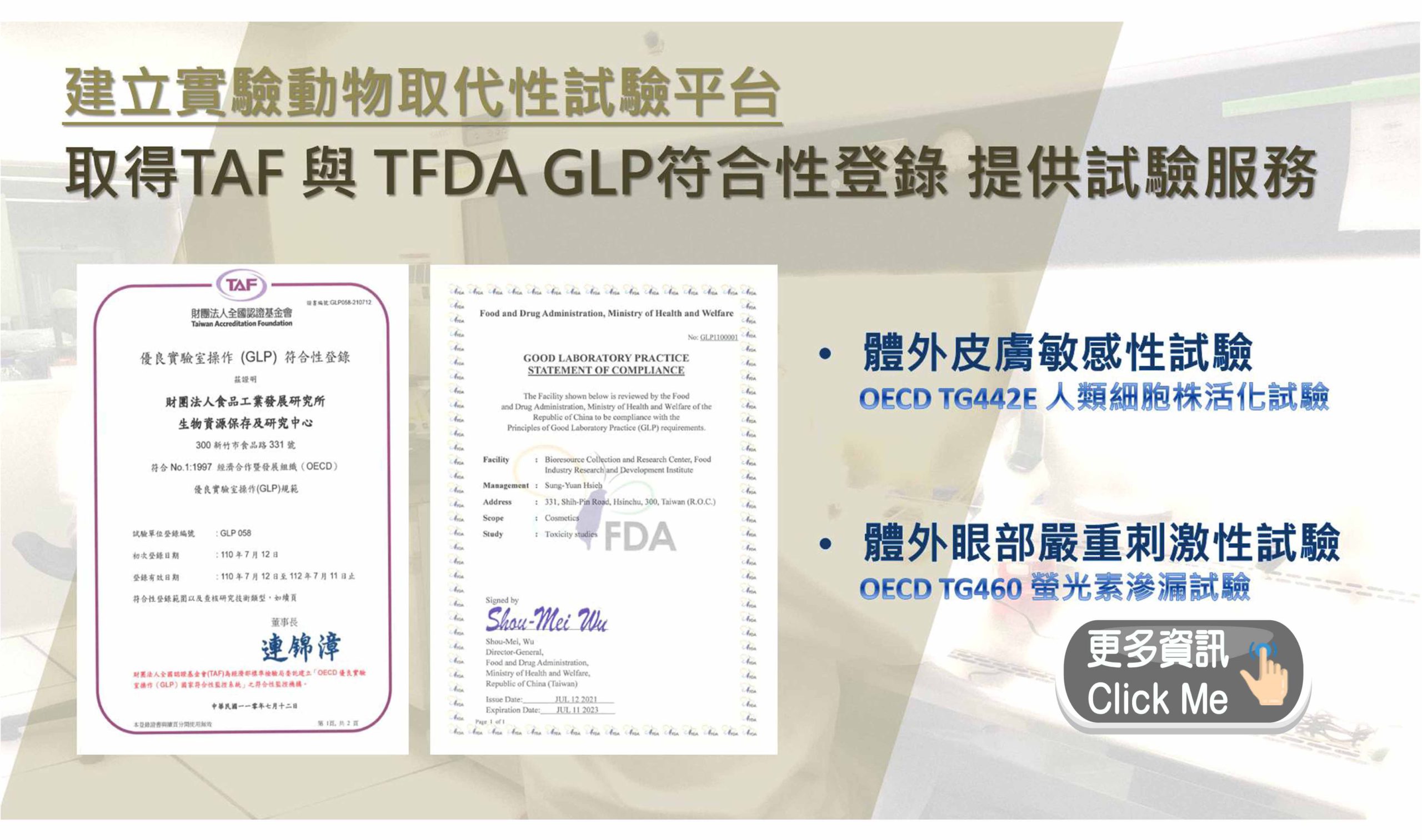
委託試驗
符合國際標準及品質認證之測試試驗項目
More
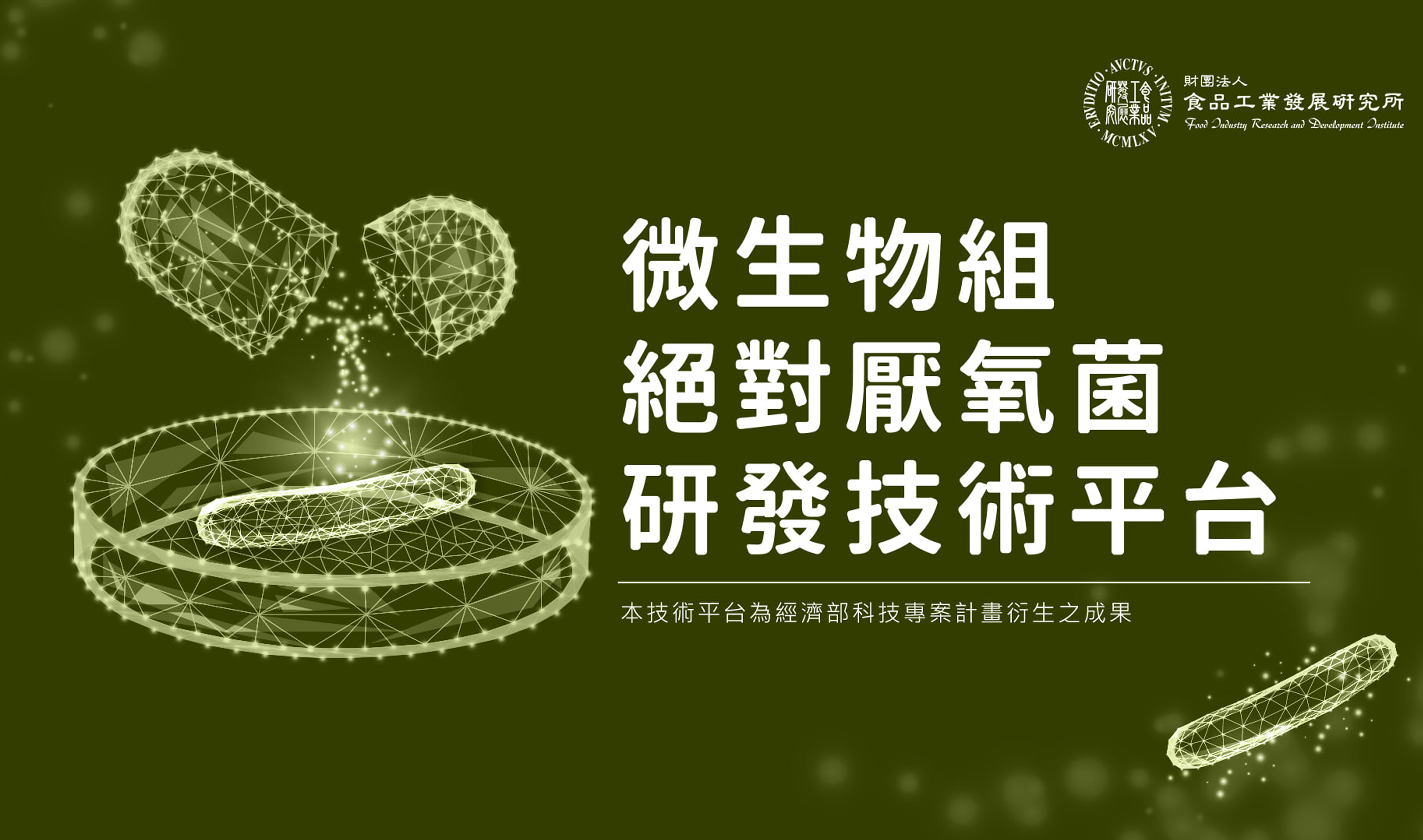
技術發展
歷年投入之研究產出及可移轉技術及專利
More
菌種鑑定
微生物及細胞鑑定、複核及客製化服務
More
菌株寄存
公開、秘密、專利及農藥微生物等之寄存
More
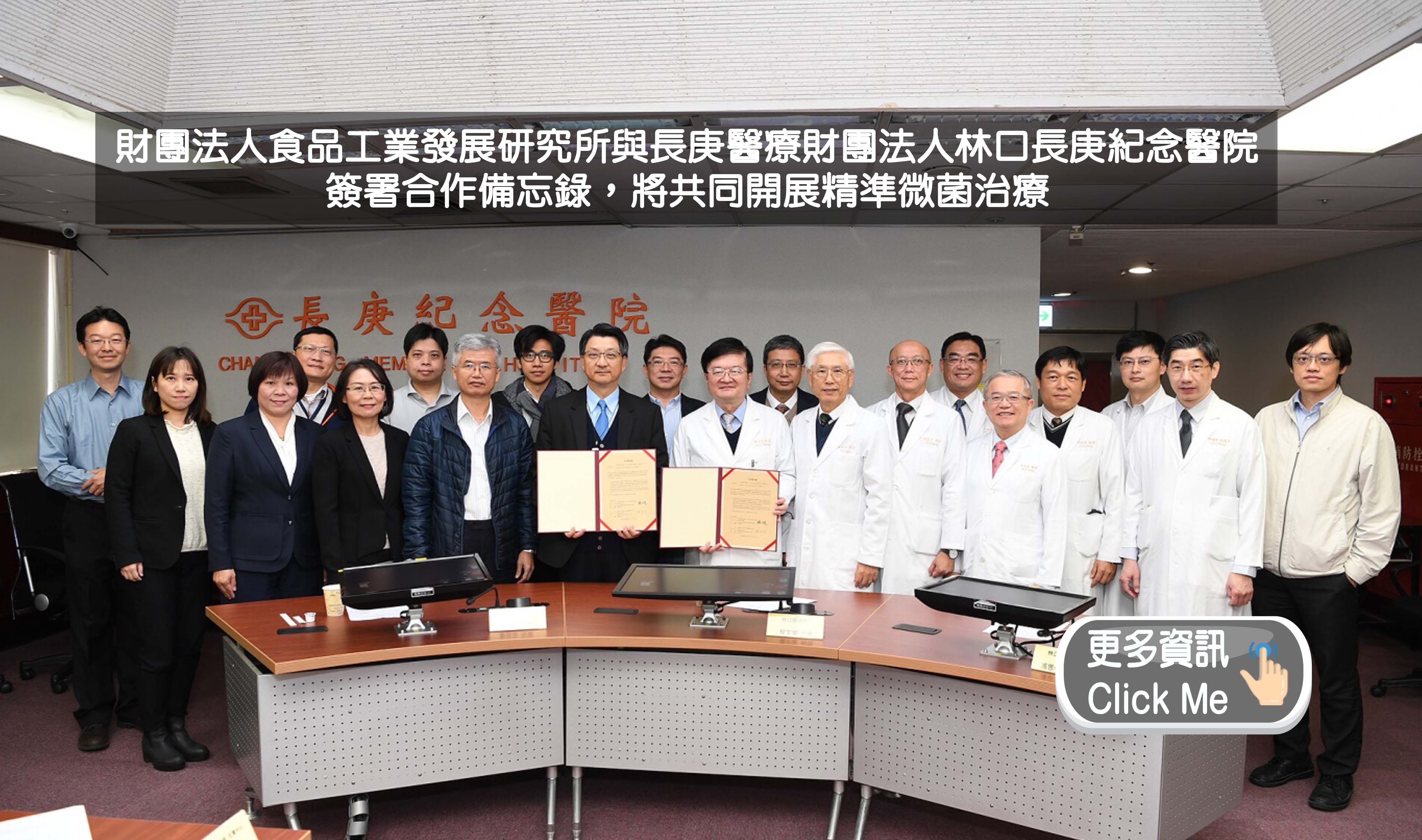
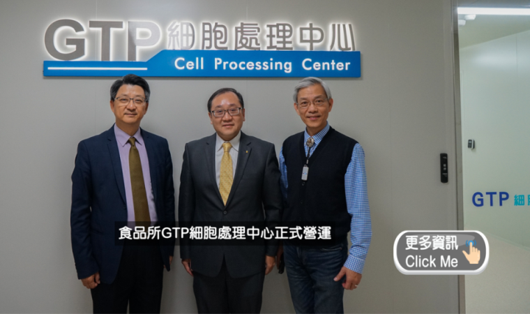
即時訊息
最新消息
網頁新知
生資中心簡訊
最新消息
2025-03-24
114年清明節連假細胞株暫停出貨一次
2025-03-20
敬邀參與食品所114年研發成果展示暨產研合作計畫說明會
2025-02-26
114年度經濟部智慧財產局指定專利生物材料之國內寄存機構為財團法人食品工業發展研究所
2025-01-09
114年農曆春節期間生資中心暫停服務公告
2024-12-19
生物資源線上目錄暫停通知
+更多資訊
網頁新知
2025-06-30
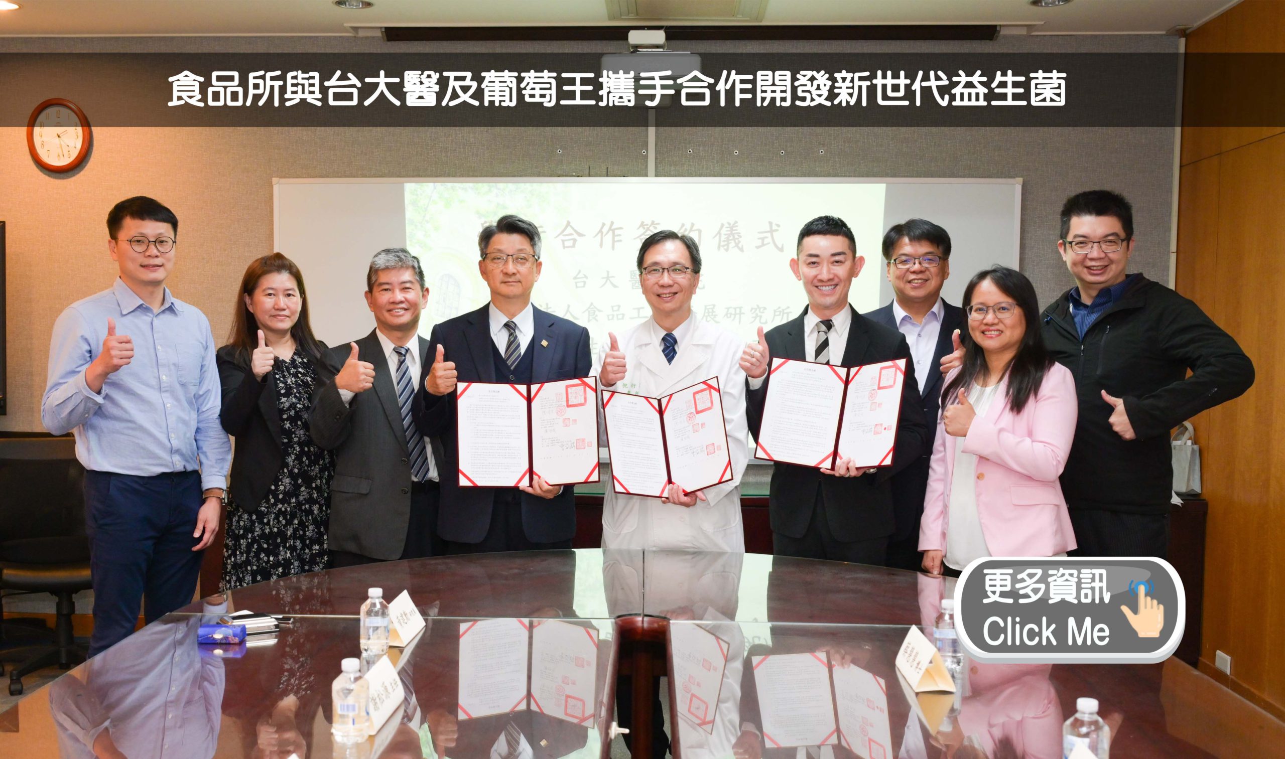
基因編輯香蕉:告別爛果宿命,解鎖全球糧食新篇章
2025-06-09
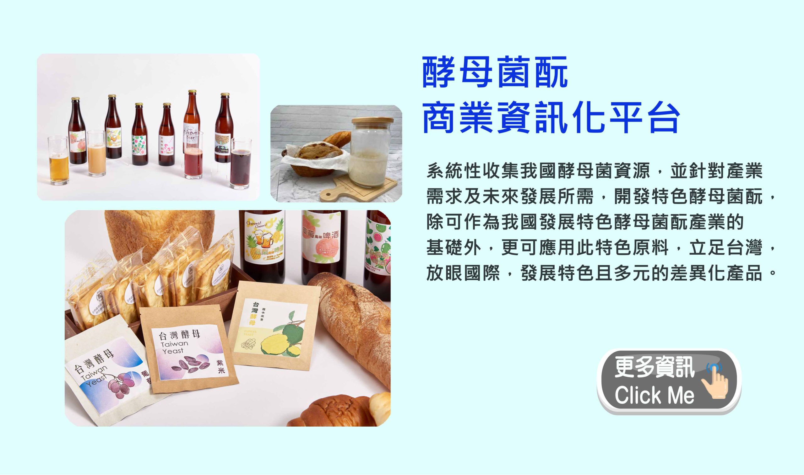
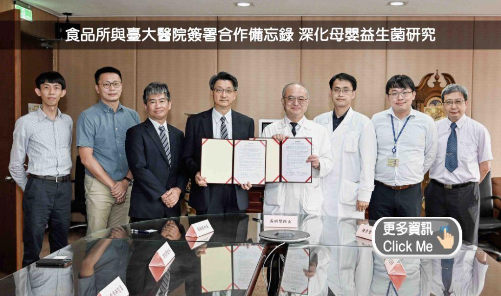
益生菌對創傷性腦損傷後神經保護與腸道菌相之效應
2025-06-09
生物樣本庫,讓世界更健康
2025-04-29
LINE的AI助理服務 -「AI小幫手」
2025-04-29
腫瘤生態系中的隱藏玩家—真菌
+更多資訊
生資中心簡訊
第136期簡訊
第135期簡訊
第134期簡訊
第133期簡訊
第132期簡訊
+更多資訊
推薦影音
Menu
首頁
關於生資中心
中心簡介
中心組織
中心沿革
創新研發與應用
生物材料寄存及分讓辦法
公正性聲明
關鍵技術
生物資源之培養與保存技術
生物資源之開發與應用技術
對外服務
生物資源提供
生物資源引進
委託試驗
菌種鑑定與人類細胞複核
寄存服務
秘密寄存
公開寄存
專利寄存
農藥微生物種源寄存
委託代訓
最新消息 & 網頁新知
出版品
生資中心簡訊
其他出版品
聯絡我們
FAQs
相關聯結
生物資源線上目錄
BCRC客戶服務系統
微生物條碼資料庫系統
BCRC小教室
專利生物材料服務e平台
台灣微生物知識網
國家衛生研究院細胞庫
產業服務平台
TSCB台灣幹細胞庫
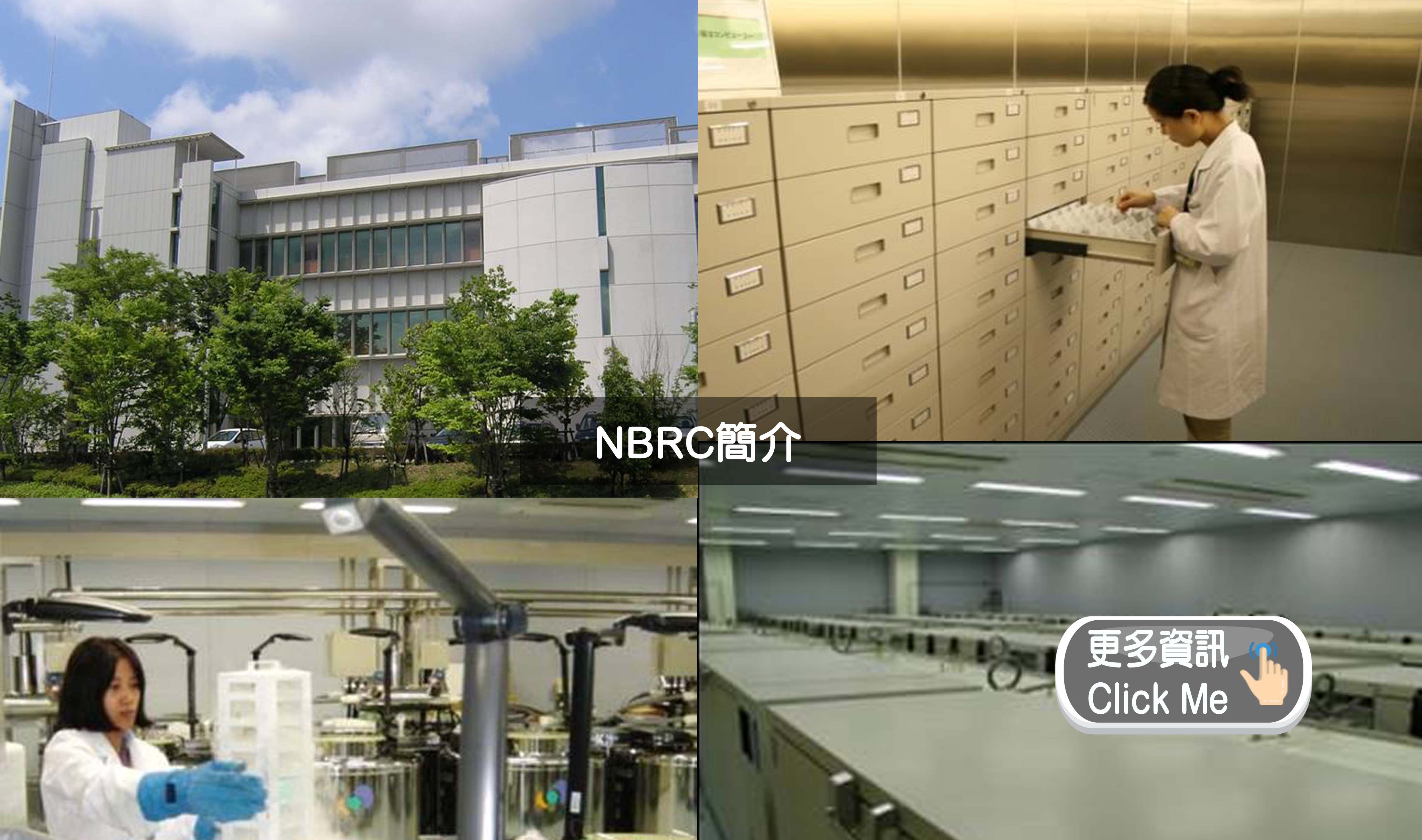
新資源
NBRC-BCRC 國際合作專區
同仁專區
BCRC工作流程管理系統
生物資源管理系統
最新消息
2025-03-24
114年清明節連假細胞株暫停出貨一次
2025-03-20
敬邀參與食品所114年研發成果展示暨產研合作計畫說明會
+更多資訊
網頁新知
2025-06-30
基因編輯香蕉:告別爛果宿命,解鎖全球糧食新篇章
2025-06-09
益生菌對創傷性腦損傷後神經保護與腸道菌相之效應
+更多資訊
生資中心簡訊
第136期簡訊
第135期簡訊
+更多資訊
對外服務
菌株購買
多樣、本土、新穎性及應用性生物資源
更多資訊
委託試驗
符合國際標準及品質認證之測試試驗項目
更多資訊
技術發展
歷年投入之研究產出及可移轉技術及專利
更多資訊
菌種鑑定
微生物及細胞鑑定、複核及客製化服務
更多資訊
菌株寄存
公開、秘密、專利及農藥微生物等之寄存
更多資訊
主題連結